Journal of Animal Health and Production
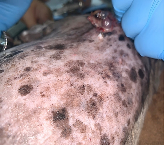
Generalized brown-greyish and blackish macules of varying sizes with irregular borders.
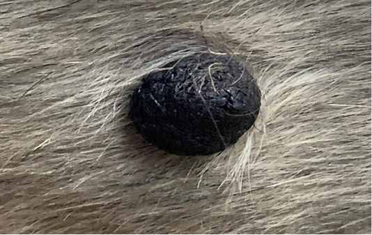
Focal and soft black nodules with a smooth surface of 1 cm in size observed at the right lateral trunk region.
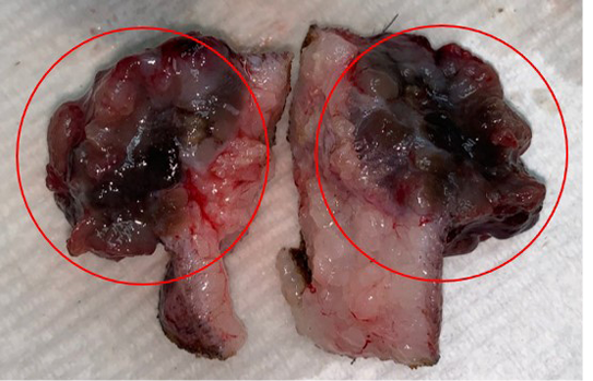
Brownish-black mass with cauliflower-like appearance (circles).
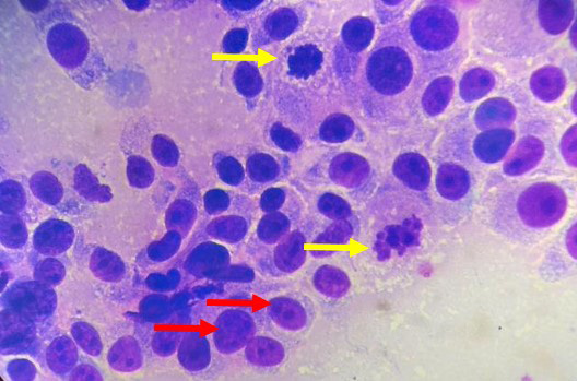
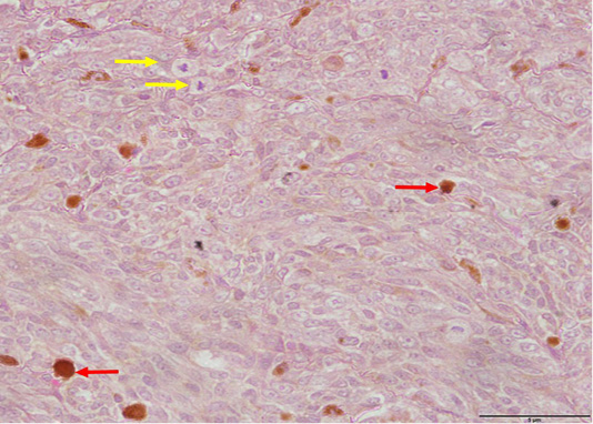
Pleomorphism characterized by a mixture of spindle and round cells and presence of multiple nucleoli (red arrows), mitotic figures (yellow arrows) and intracytoplasmic melanin granules (H and E, 1000X).
Pleomorphism with spindle and round cells, anisokaryosis and mitotic figures (yellow arrows) as well as intracytoplasmic melanin pigments observed within the dermis (red arrows) (H and E, 400X).
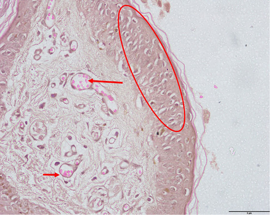
Pagetoid melanocytosis (circle) and evidences of angiogenesis (arrows) (H and E, 400X).


