Research Journal for Veterinary Practitioners
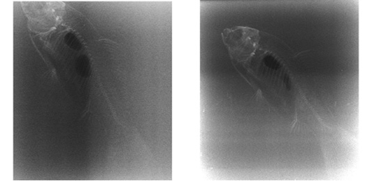
Laterolateral horizontal projection of the goldfish (Carassius auratus, Linnaeus 1758) just before (a) and after (b) puncture of the posterior camera.
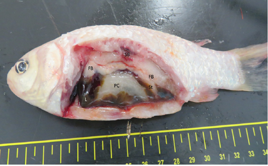
Morphology and location of the coelomic organs of a goldfish (Carassius auratus, Linnaeus 1758) after incision (SB – swim bladder; FB – fat bodies; St, stomach)
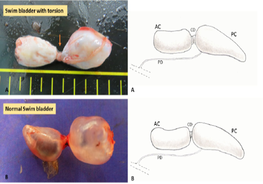
In A swim bladder torsion, the point (arrow) where the torsion happened, and in B a normal swim bladder (anterior chamber -AC, posterior chamber -PC, communicating duct -CD and pneumatic duct -PD). On the left a schematic representation of the torsion (A) and normal swim bladder (B).
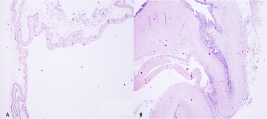
Anterior chamber presented a single layer of flattened epithelial cells, with a prominent basal lamina below the epithelial cells and lamina propria containing collagen fibrils, elastic fibres, and fibroblasts. In B section in the point where the torsion occurred. Many cells were degraded due to the rapid decomposition of the corpse (H&E, 40x).


