Research Journal for Veterinary Practitioners
Case Report
Res. J. Vet. Pract. 4(2): 25-29
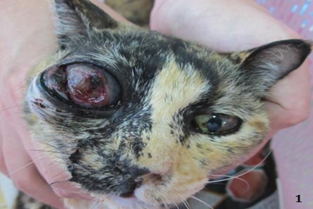
Figure 1
DSH cat presented with intraocular and retrobulbar mass affecting the right eye
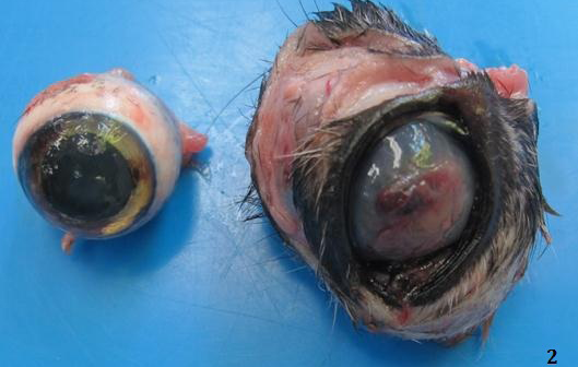
Figure 2
Right intraocular mass with ulcerative keratitis
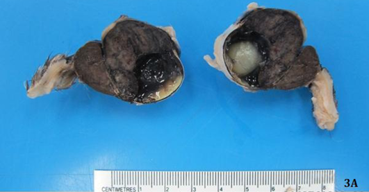
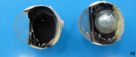
Figure 3
Firm large brown to black retrobulbar and intraocular mass effacing both anterior and posterior chambers of the right eye (Figure 3A) compared to normal left eye (Figure 3B)
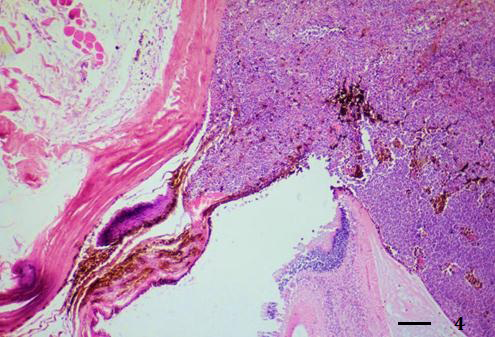
Figure 4
Extensive melanoma occupied iris and posterior chamber (H&E stain, bar = 250 µm)
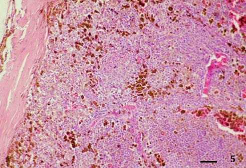
Figure 5
Diffuse solid nests of tumor cells in posterior chamber (H&E stain, bar = 100 µm)
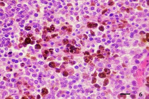
Figure 6
Neoplastic cells had large epithelioid shape and contained intracytoplasmic brown black melanin pigments (H&E stain, bar = 25 µm)
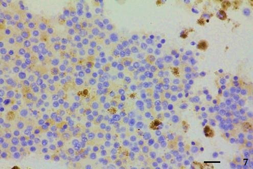
Figure 7
Tumor cells showed intense cytoplasmic Melan-A protein expression (IHC; Envision polymer, counterstained with Mayer’s Hematoxylin, bar = 25 µm)


