Advances in Animal and Veterinary Sciences
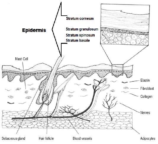
Diagram shows the epidermis with stratum corneum, stratum granulosum, stratum spinosum and stratum basale. Also dermis with its structures in the cutaneous dog.
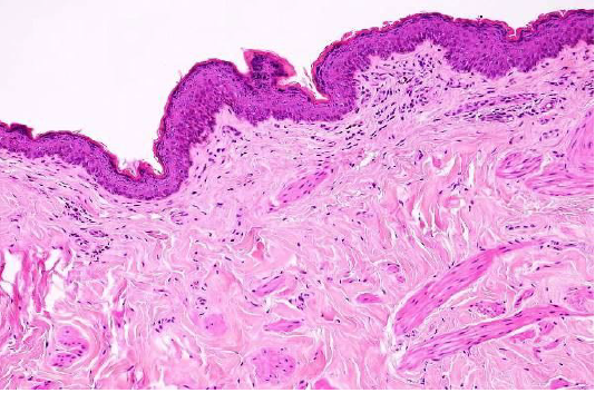
Showed the normal structure of epidermis and dermis in the cutaneous dog (H&E×40) (Dirrig et al. 2016).
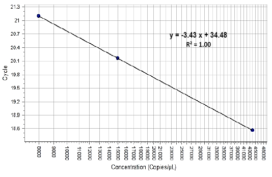
Showed the standard curve
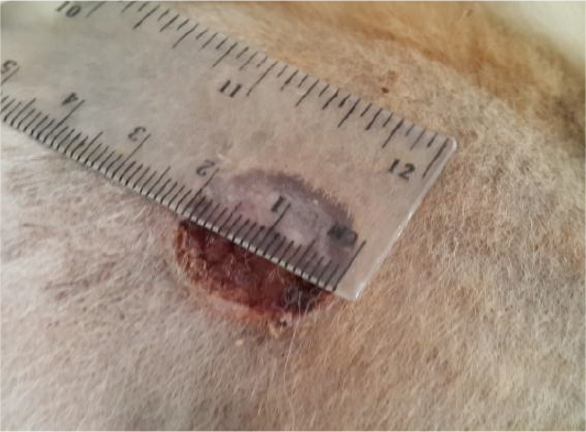
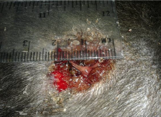
Showed like volcano orifice protrusion
Showed the suppurative exudate formation
Showed the epidermis (white ruler) stratum corneum (A), stratum granulosum (B), stratum spinosum (C) and stratum basale (D). Dermis (black ruler) blood vessel (black arrow). The preparation was by (H&E×40).
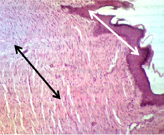
Showed the epidermis (white arrow) and dermis (black arrow) convert from irregular arrangement to regular pattern. The preparation was by (H&E×40).
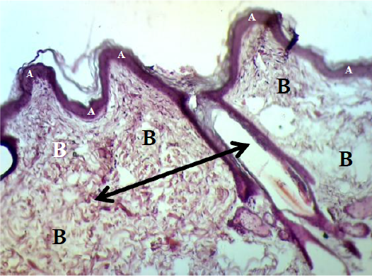
Showed the epidermis (A) consists stratum corneum, stratum granulosum, stratum spinosum and stratum basale. Dermis (B) consists big blood vessel (black arrow). The preparation was by (H&E×40).
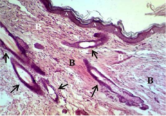
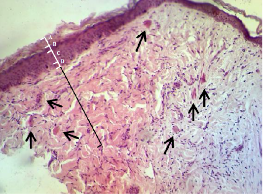
Showed the epidermis (A) consists stratum corneum, stratum granulosum, stratum spinosum and stratum basale. Dermis (B) immature to mature granulation tissue (black arrow). The preparation was by (H&E×40).


