Advances in Animal and Veterinary Sciences
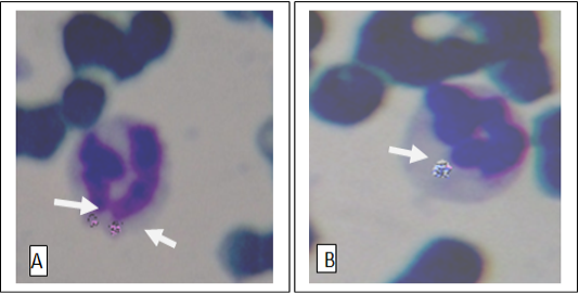
Anaplasma phagocytophilia organisms in a neutrophil (A) and Monocyte (B) Buffy coat smear; Giemsa stain. Magnification, 1,000.
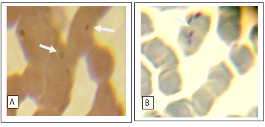
Numerous small basophilic structures of hemotropic Mycoplasma spp. organisms on the surface of the erythrocytes attached to cell membranes singularly(A) or in clustered (B). Peripheral blood smear; Giemsa stain. Magnification, 1,000.
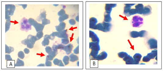
Giemsa stain light micrograph (Magnification, 1,000) of horse blood smear, co-infection of hemotropic Mycoplasma spp. and A. phagocytophila
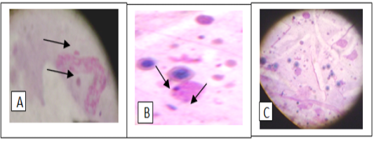
Giemsa stain of tick hemolymph cells infected with Anaplasma phagocytophilium (A), hemotropic Mycoplasma spp. (B) and co- infection of both bacteria (C)


