Advances in Animal and Veterinary Sciences
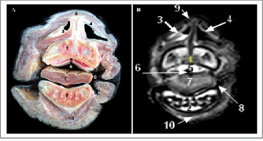
Cross sectional anatomy (Panel A) and MR image (Panel B) of horse head at the level of upper and lower incisors. 1) Upper incisors teeth; 2) Lower incisors teeth; 3) plate of alar cartilage; 4) Horn of alar cartilage; 5) Hard palate; 6) Oral cavity proper; 7) Apex of tongue; 8) Labial vestibule; 9) Apical naris dilatator muscle; 10) Mentalis muscle.
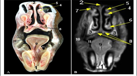
Cross sectional anatomy (Panel A) and MR image (Panel B) of horse head at the level of body of mandible. 1) Body of mandible; 2) Nasal bone; 3) Maxilla bone; 4) medial accessory nasal cartilage at Alar fold; 5) Straight fold; 6) Basal fold; 7) False nostril (Nasal diverticulum); 8) Vomeronasal organ; 9) Tongue; 10) Buccinators muscle; 11) Sublingual recess.
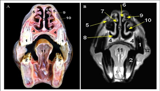
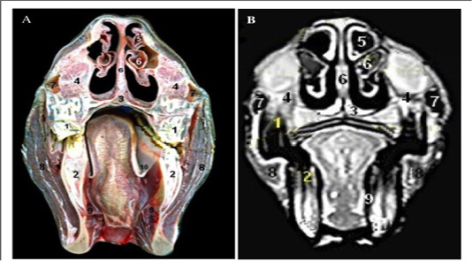
Cross sectional anatomy (Panel A) and MR image (Panel B) of horse head at the level of first and second premolar teeth. 1) Upper first and second premolar teeth; 2) Lower first and second premolar teeth; 3) Cartilaginous part of Nasal septum; 4) maxilla bone; 5) Nasal bone; 6) Dorsal nasal meatus; 7) Middle nasal meatus; 8) Ventral nasal meatus; 9) Dorsal nasal concha; 10) Ventral concha1 bulla; 11) Body of tongue; 12) Buccinators muscle.
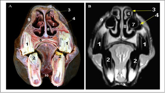
Cross sectional anatomy (Panel A) and MR image (Panel B) of horse head at the level of the third premolar tooth. 1) Upper third premolar tooth; 2) Lower third premolar tooth; 3) Dorsal turbinate crest of Nasal bone; 4) Ventral turbinate crest of maxilla bone; 5) Palatine process of Maxilla; 6) Dorsal nasal concha; 7) Ventral nasal concha (bulla); 8) Common nasal meatus; 9) Superior labii levator muscle; 10) Geniohyoid muscle.
Cross sectional anatomy (Panel A) and MR image (Panel B) of horse head at the level of the molar teeth. 1) Upper molar teeth; 2) Lower molar teeth; 3) Vomer; 4) Maxilla bone; 5) Dorsal conchal bulla; 6) Ventral conchal sinus; 7) Rostral maxillary sinus; 8) Masseter muscle; 9) Pteyrgoid muscle (medial part); 10) Palatoglossal fold.
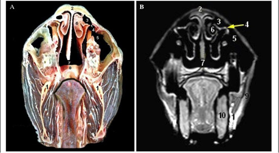
Cross sectional anatomy (Panel A) and MR image (Panel B) of horse head at the level of the diastema. 1) Ramus of mandible; 2) Nasal bone; 3) Concho frontal sinus (rostral part); 4) Fronto-maxillary opening; 5) Caudal maxillary sinus; 6) Dorsal nasal sinus (Caudal end of dorsal nasal concha); 7) Nasopharynx; 8) Soft palate; 9) Masseter muscle; 10) Pteyrgoid muscle (medial part).
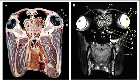
Cross sectional anatomy (Panel A) and MR image (Panel B) of horse head at the level of the perpendicular plate of ethmoidal bone.1) Perpendicular plate of ethmoidal bone; 2) Ethmoid labyrinth (ethmoid conchae); 3) Vomer bone; 4) Maxilla; 5) Mandible; 6) Zygomatic arch; 7) Orbit; 8) Conchofrontal sinus (caudal part); 9) Sphenopalatine sinus; 10) Middle conchal sinus; 11) Septum of frontal sinus; 12) Nasopharynx; 13) Soft palate; 14) cornea; 15) Sclera; 16) Lens; 17) Retina; 18) Extra-ocular muscles.


