Advances in Animal and Veterinary Sciences
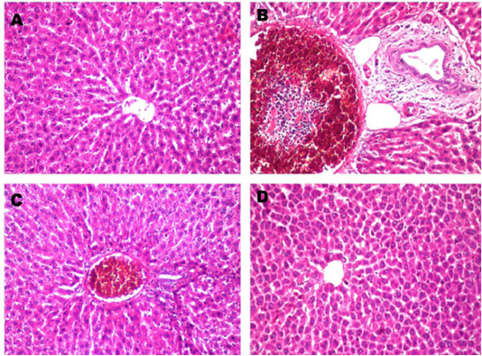
Histopathological changes in liver after treated with paracetamol and/or Ocimum basilicum. A: Normal liver section from control rat shows no histopathological alteration and the normal histological structure of the central vein and surrounding hepatocytes in the parenchyma (X10). B: Paracetamol treated rats showed sever congestion and dilatation in the portal vein associated with inflammatory cells infiltration and oedema in the periductal tissue surrounding the cystic dilated bile ducts (X20). C: Paracetamol + Ocimum basilicum (200 mg/kg bwt) treated rats, showed congestion in the portal vein associated with few inflammatory cells infiltration surrounding the bile ducts (X10) and D: Paracetamol + Ocimum basilicum (400 mg/kg bwt) treated rats, there was no histopathological alteration were recorded (H and E).
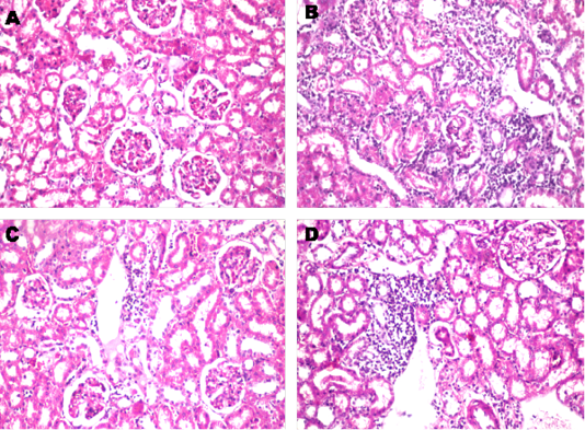
Histopathological changes in renal cortical tubules after treated with paracetamol and/or Ocimum basilicum. A: Kidney section from control rat shows the normal histological structure of the glomeruli and tubules at the cortex (X10). B: Paracetamol treated rat shows, Massive inflammatory cells infiltration was detected in between the degenerated renal tubules at the cortex (X10). C Paracetamol + Ocimum basilicum (200 mg/kg bwt) treated rats, showed focal inflammatory cells infiltration was detected in the perivascular area surrounding the cortical blood vessels (X10) and D: Paracetamol + Ocimum basilicum (400 mg/kg bwt) treated rats, focal inflammatory cells infiltration was detected in between the tubules at the cortex (H and E).


