Advances in Animal and Veterinary Sciences
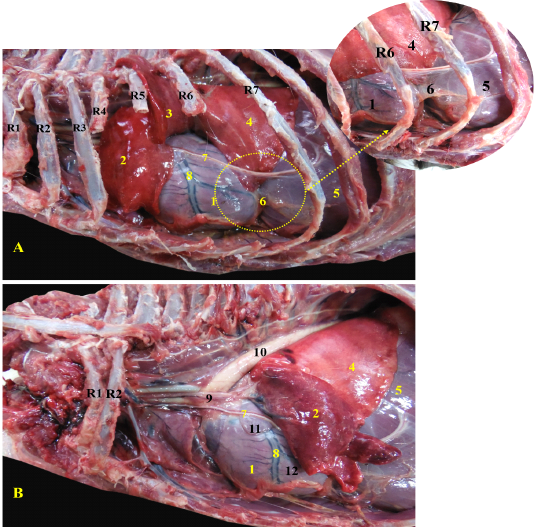
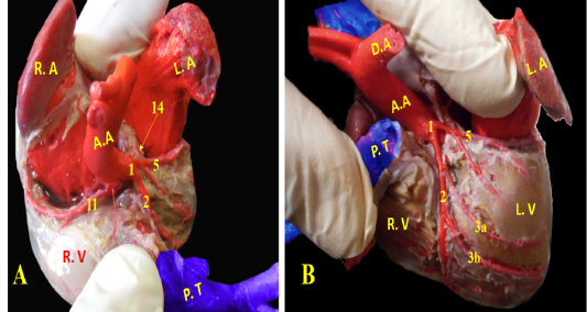
Showing the heart of red fox insitu A- showing the heart insitu after rib reflection, B - showing the heart after reflection of the left cranial lobe of lung.
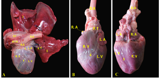
Showing the separated heart of red fox. A- showing the heart and lung separate with pericardium, B- showing the left side of the heart without pericardium, C- showing the right side of the heart without pericardium.
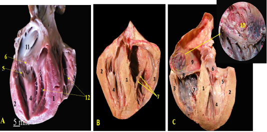
A, B, C showing the sagittal sections of the heart of red fox. With scale bar= 5mm, 1-Interventricular septum, 2- wall of left ventricle, 3- wall of right ventricle, 4- papillary muscle of left ventricle, 5- chorda tendineae of mitral valve, 6- valvular leaflet of mitral valve, 7- papillary muscle of right ventricle, 8- valvular leaflet of tricuspid valve, 9- right atrium, 10- pectinate muscle of right atrium, 11-left atrium, 12- trabeculae cornae
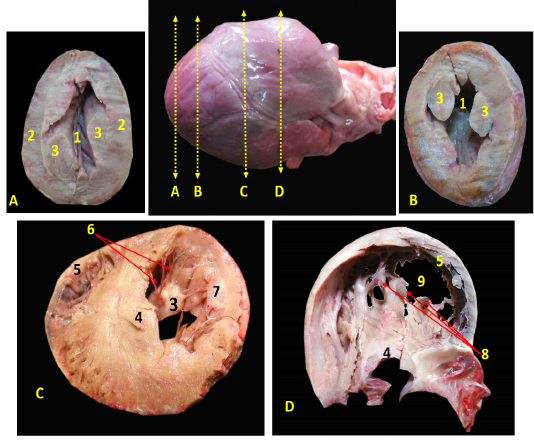
Showing the cross sections of the heart of red fox at different levels.
Showing coronary arteries, A- showing the origin of coronary arteries, B- showing the main branches of left coronary.
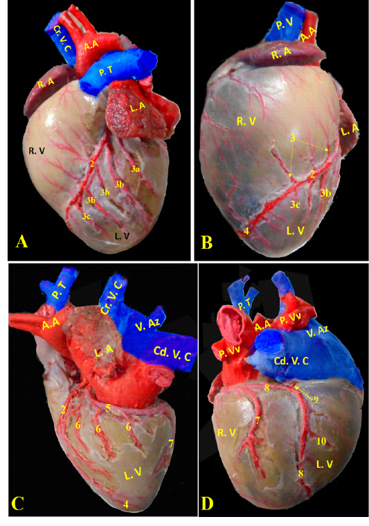
Showing division of left coronary arteries, A, B- showing the branches of left paraconal artery. C, D- showing the branches of left circumflex artery.
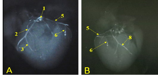
Showing radiographic images for division of the left coronary arteries, A- showing the left coronary artery. B- showing the branches of left circumflex artery.
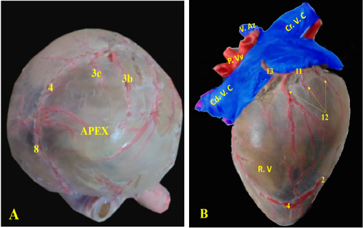
A-Showing the apex of the heart., B-showing the division of right coronary artery.


