Advances in Animal and Veterinary Sciences
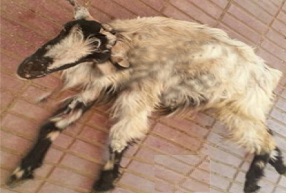
A doe with PT and exhibit nervous signs include incoordination, star-gazing, tremors, and convulsions
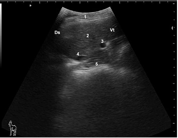
Ultrasonogram of hepatic parenchyma and hepatic blood vessels in a healthy late pregnant doe viewed from the 10th ICS on the right side
1: Lateral abdominal wall; 2: Liver parenchyma; 3: Portalvein; 4: Caudal vena cave; 5: Omasum; Ds: Dorsal; Vt: Ventral
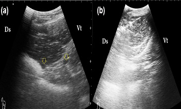
Ultrasonogram of liver in PT does
A: a good prognosis case; the liver appears more echogenic than the normal with visible hepatic vessels (arrows); B: a bad prognosis case: the liver appears more echogenic than the normal with invisible hepatic vessels; Ds: Dorsal; Vt: Ventral
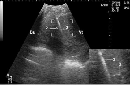
Hepatic biopsy in a doe, notice the needle is clearly visible as hyper-echoic structure (zoom area) within the hepatic parenchyma
1: Liver parenchyma; 2: Biopsy needle; 3: Portal vein; Ds: Dorsal; Vt: Ventral
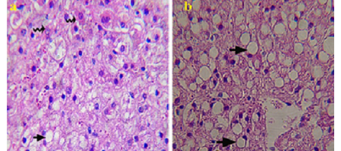
Histopathological picture of liver specimen obtained by biopsy from PT does
A: showing mild macro vesicular fatty change (arrow) and numerous normal hepatocytes (zigzag arrow) (good prognosis); B: showing severe vesicular fatty change (arrow) and less normal hepatocytes (bad prognosis). H & E Χ 1200
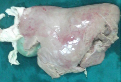
Liver of severely affected PT doe showing fatty change at necropsy examination


