Advances in Animal and Veterinary Sciences
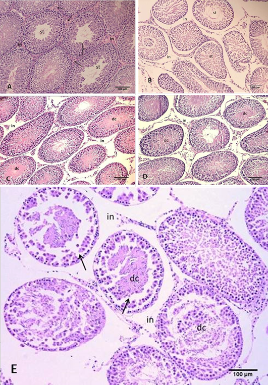
Section of the testis
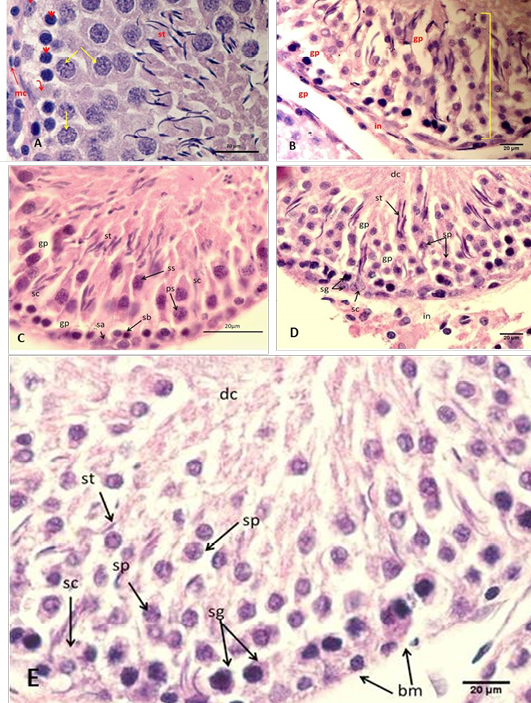
Section of the testis
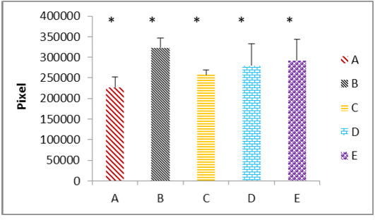
The immunohistochemical analysis of testicular tissues of the five groups
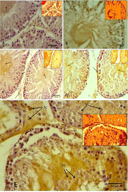
The immunohistochemical micrography of testicular tissues
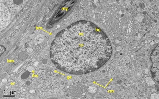
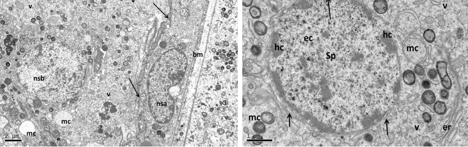
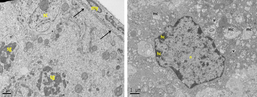
Electron micrograph (TEM) of rat testes from fresh control group (A) Showing (Left) normal cellular components
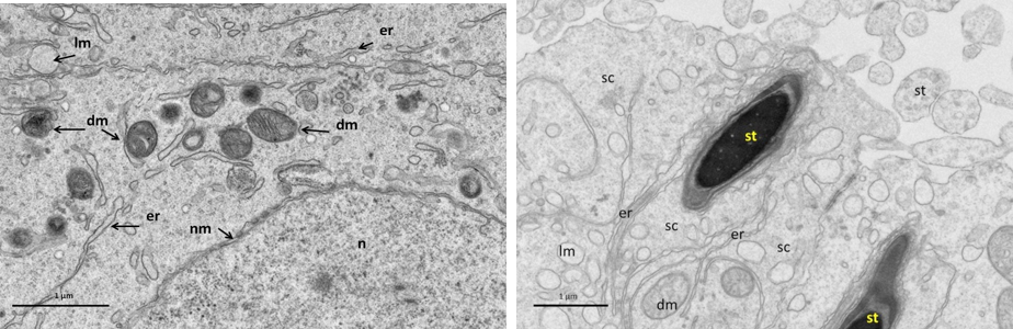
Electron micrograph (TEM) of Sertoli cell from fresh control group (A)
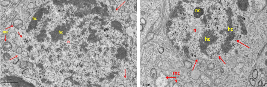
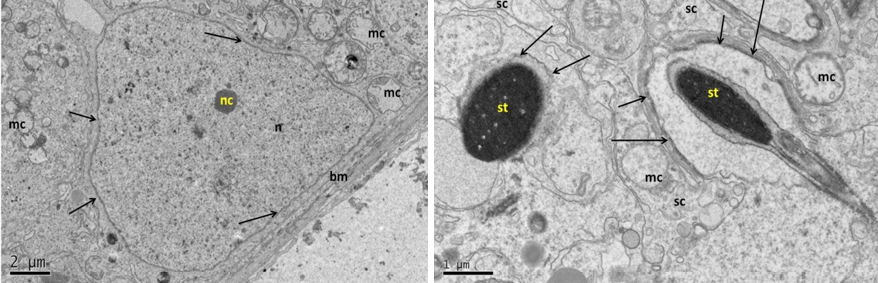
Electron micrograph (TEM) of rat testes from group (B) cryopreserved with freezing media only showing sever cellular changes
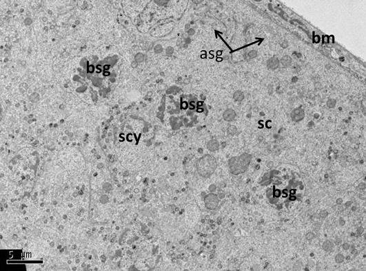
Electron micrograph (TEM) of spermatogonia (Left) and spermatocyte (Right) from group (B) cryopreserved with freezing media
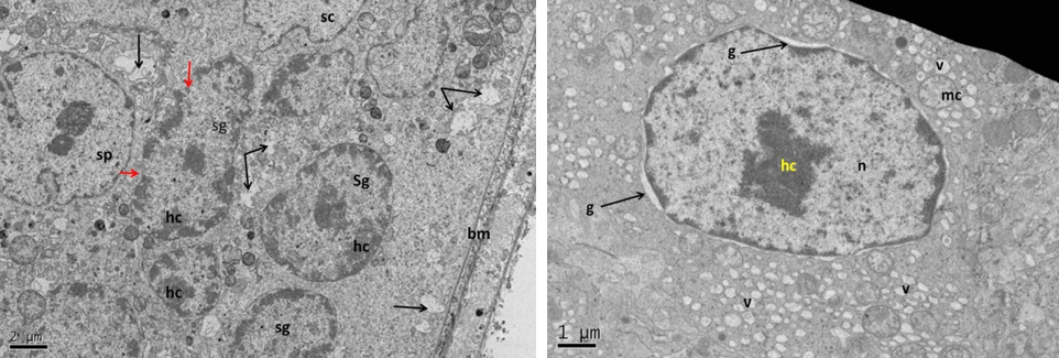
Electron micrograph (TEM) of rat testes from group (C) cryopreserved with DMSO showing (Left) Light cellular changes
Electron micrograph (TEM) of Sertoli cell from group (C) cryopreserved with DMSO
Electron micrograph (TEM) of rat testes from group (D) cryopreserved with glycerol showing (Left) cellular changes
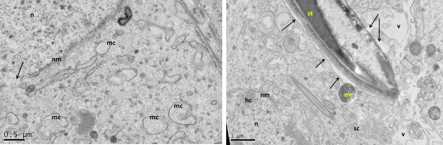
Electron micrograph (TEM) of Sertoli cell from group (D) cryopreserved with glycerol
Electron micrograph (TEM) of rat testes from group (E) cryopreserved with 1, 2 prOH showing (Left) cellular changes


