Advances in Animal and Veterinary Sciences
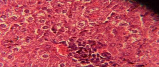
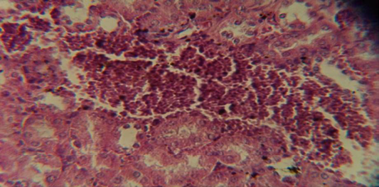
Histological section in liver of infected group showing multiple focal granulamatous lesion in liver parenchyma especially around central vein consist of aggregation of mononuclear cells
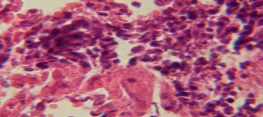
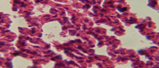
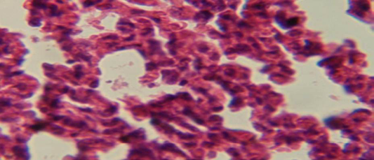
Histological section in lung of infected group showing large granulomatous lesion consist of aggregation of macrophages in lung parenchyma
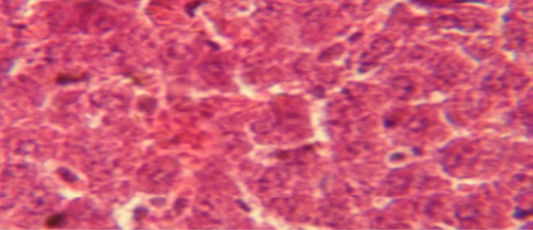
Histological section in liver of treatment group with ethambutol + extract showing aggregation of mononuclaur cell in liver parenchyma and proliferation of kupffer cell
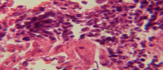
Histological section in lung of infected group showing large granulamatous lesion consist of aggregation of macrophages in lung parenchyma
Histological section of lung of treatment group with ethambutol revealed congestion of blood vessel with few inflammatory cell in their lumen
Histological section of lung of treatment group with ethambutol + extract showing aggregation of mononuclear cell in the interalveolar septa
Histological section in kidney of infected group showing congestion of blood vessel with infiltration of inflammatory cell in their lumen
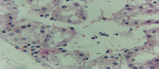
Histological section of kidney of treatment group with ethambutol revealed cellular degeneration of epithelial cell lining renal tubule and neutrophil infiltration in the interstitial tissue
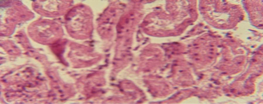
Histological section of kidney of treatment group with ethambutol + extract revealed cellular degeneration of epithelial cell lining renal tubule


