Advances in Animal and Veterinary Sciences
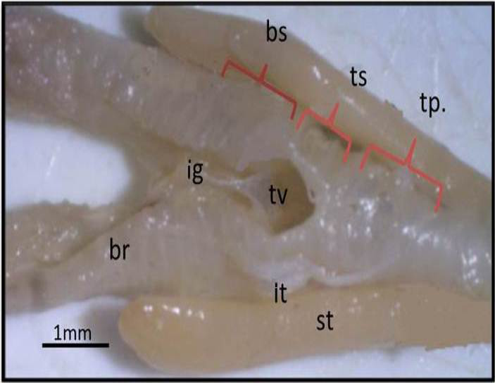
Macrograph of black francolin syrinx (ventral view): showing the parts of syrinx that formed by: tympanum (tp); trachea-syringeal part (ts); broncho-Syringeal part (bs); triangular voice (tv); ligament (ig); sternotrachilis (st); lateral trachealis (lt); primary bronchi (br).
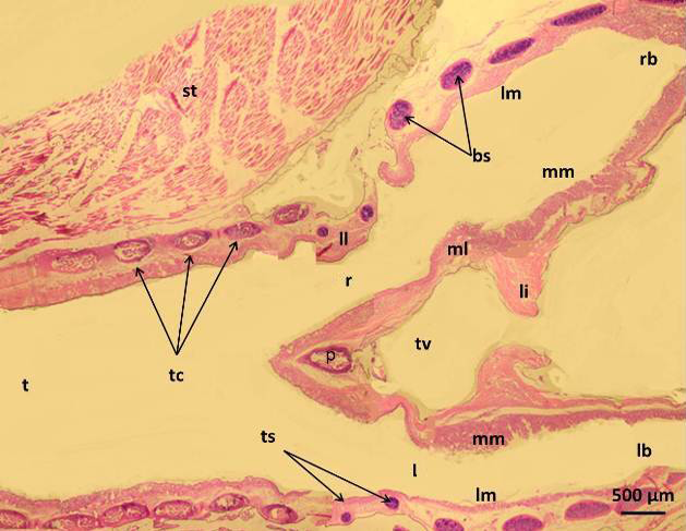
Micrograph of syrinx of male black francolin showing: trachea (t); tympanum cartilages (tc); trachea-syringeal cartilages (ts); pessulus (p); right passage (r); left passage (l); lateral labium (ll); medial labium (ml); lateral vibrating membrane (lm); medial vibrating membrane (mm); triangular voice (tv); sternotrachealis (st): ligament (li). H&E.

Macrograph of syrinx (dorsal view) of black francolin: showing tympanum which formed by (1, 2, 3, yellow color) C shaped cartilages; trachea-syringeal part which formed by (1, 2, 3, black color) C shaped cartilages; pessulus (p); ligament (lig.) Alcian blue-Alizarin red technique.
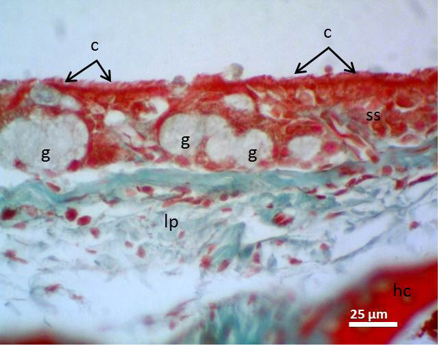
Micrograph of syrinx epithelium of black francolin. Whereas, cilia (c); pseudostratified ciliated columnar epithelia (ss); aggregation of goblet cells (intra epithelia mucous glands) (g); lamina propria (lp); hyaline cartilage (hc). Massson’s trichrom stain.
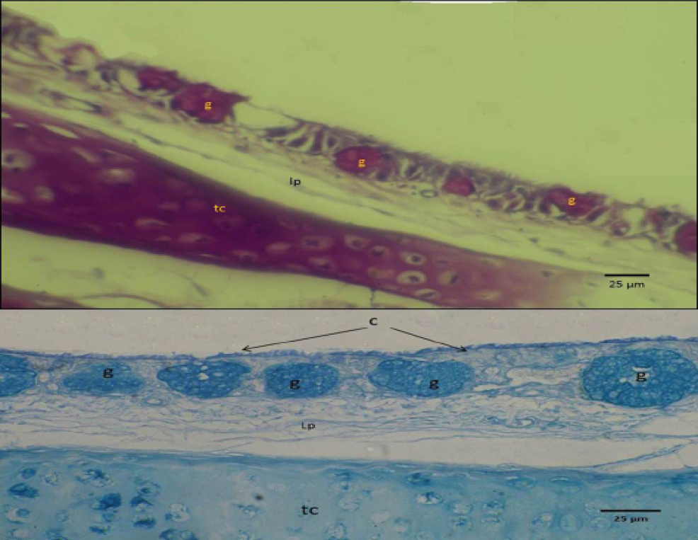
Micrograph of syrinx epithelium of black francolin. Showing mucin reaction of the mucous epithelia (upper with PAS) and (lower with Alcian blue stan), whearas, trachea-syringeal part (ts); aggregation of goblet cells (intra epithelia mucous glands) (g); lamina propria (lp).
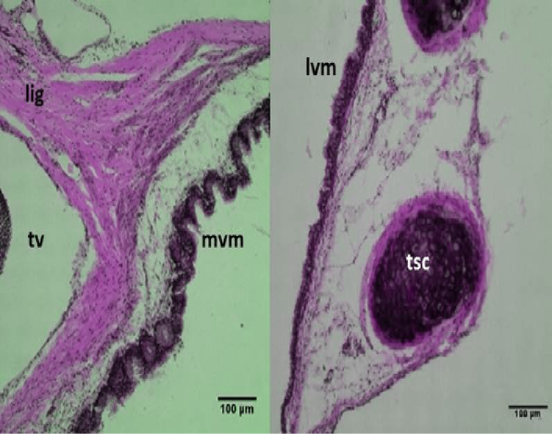
Micrograph: Medial vibrating membranes (mvm) notice the folded; lateral vibrating membranes (lvm); tracheo-syringeal cartilages (tsc); triangular voice (tv); sternotrachealis ligament (lig). (Combined Aldehyde Fuchsin-Alcian blue method).
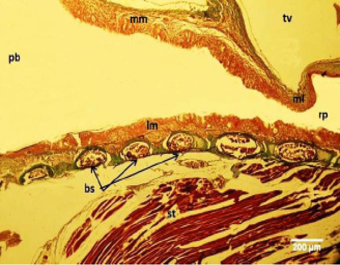
Micrograph of male black francolin syrinx. Showing ossification of the syringeal cartilages (bc); pseudostratified ciliated columnar epithelia lining the lateral vibrating membrane (lm) and medial vibrating membrane (mm); right syringeal passage (rp); primary bronchi (pb); triangular voice (tv); sternotrachealis (st). Massson’s trichrom stain.
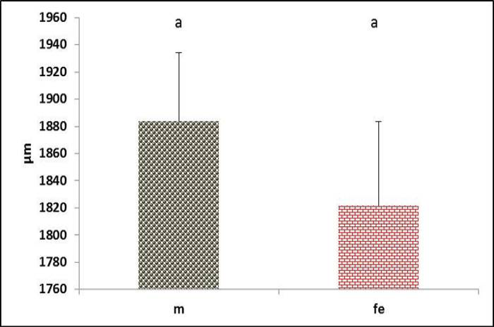
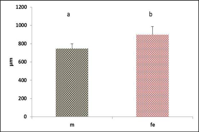
Tympanum diameter in both male and female black francolin. Values represent means±SD. Different letters means significantly (P<0.05) different. Where, male (m); female (fe).
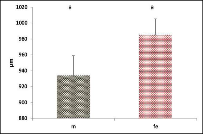
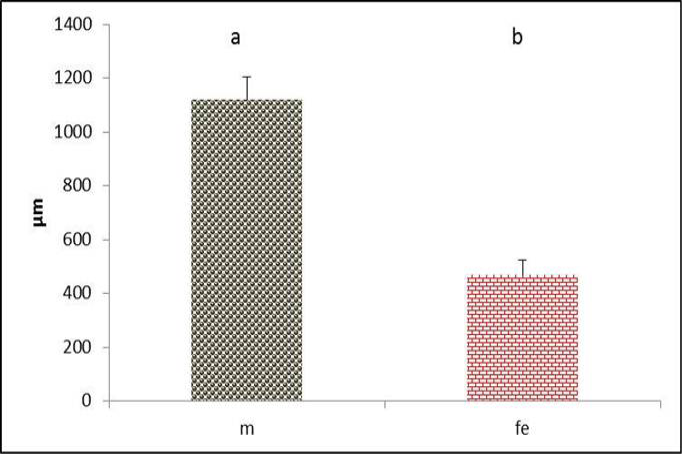
Diameter of the right syringeal passage in both male and female black francolin. Values represent means±SD. Different letters means significantly (P<0.05) different. Where, male (m); female (fe).
Diameter of the left syringeal passage in both male and female black francolin. Values represent means±SD. Different letters means significantly (P<0.05) different. Where, male (m); female (fe).
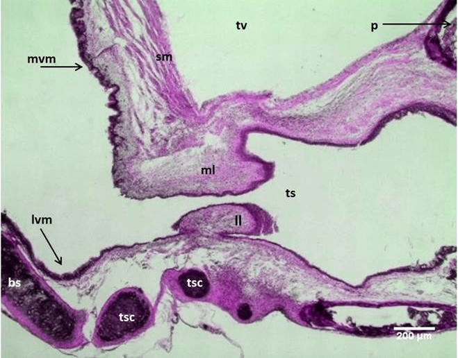
Micrograph: Longitudinal section of the syrinx in the male black francolin showing: medial labia (ml); lateral labia (ll); lateral vibrating membranes (lvm); medial vibrating membranes (mvm); tracheo-syringeal part (ts); tracheo-syringeal cartilages (tsc); triangular voice (tv); syringeal muscle (sm); broncheo-syringeal parts (bs). (Combined Aldehyde Fuchsin-Alcian blue method).
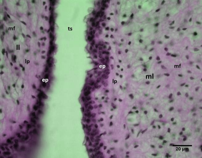
Micrograph: Syringeal labia of the male black francolin showing: medial labia (ml); lateral labia (ll); epithelium (ep); lamina propria (lp); muscle fibers (mf); passage of the tracheo-syringeal part (ts). (Combined Aldehyde Fuchsin-Alcian blue method).
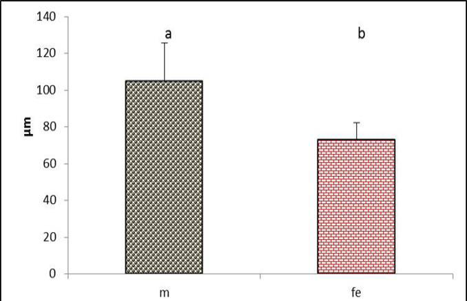
The lateral vibrating membrane thickness in the syrinx of both male and female black francolin. Values represent means±SD. Different letters means significantly (P<0.05) different. Where, male (m); female (fe).
The medial vibrating membrane thickness in the syrinx of both male and female black francolin. Values represent means±SD. Different letters means significantly (P<0.05) different. Where, male (m); female (fe).
The medial vibrating membrane thickness in the syrinx of both male and female black francolin. Values represent means±SD. Different letters means significantly (P<0.05) different. Where, male (m); female (fe).
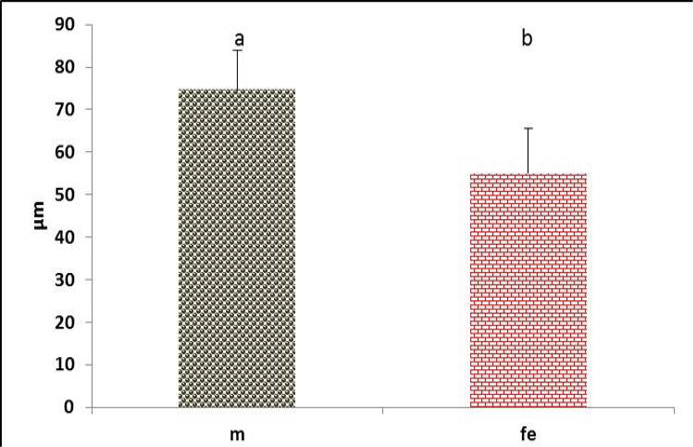
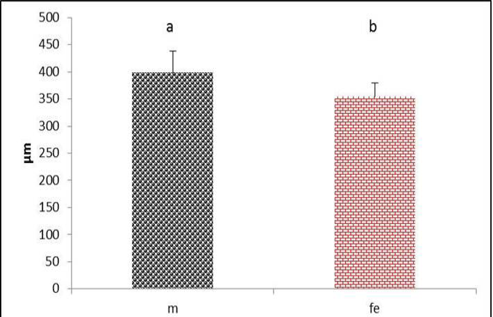
Thickness of lateral trachealis muscle in both male and female black francolin. Values represent means±SD. Different letters means significantly (P<0.05) different. Where, male (m); female (fe).
Micrograph: The Pessulus (Longitudinal section) of the syrinx in female black francolin. Notice, elongate triangle pessulus showing ossification in most of its parts. Masson´s trichrome stain.

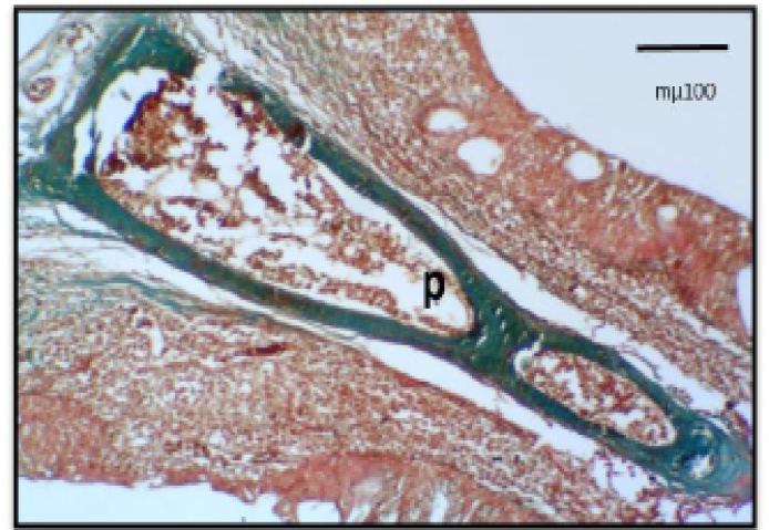
Micrograph: The Pessulus (Longitudinal section) of the syrinx in male black francolin. Notice, short wide triangle pessulus showing ossification in most of its parts. Masson´s trichrome stain.
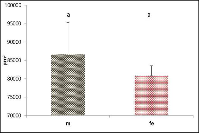
Pessulus area of syrinx in both male and female black francolin. Values represent means±SD. Different letters means significantly (P<0.05) different. Where, male (m); female (fe).
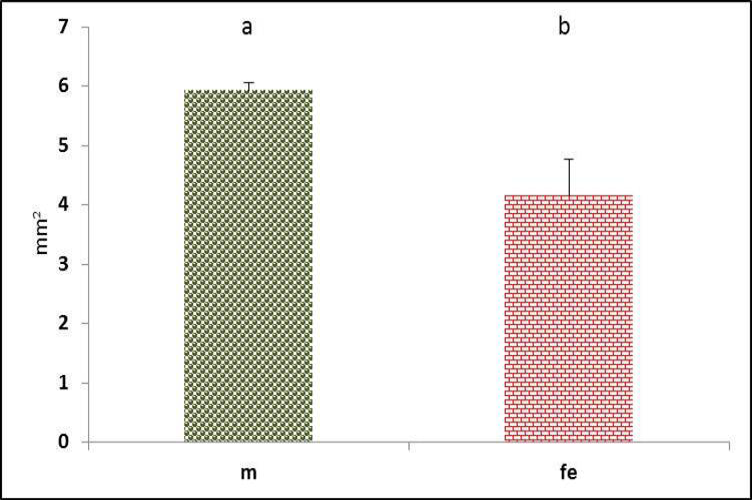
Triangular voice area of syrinx in both male and female black francolin. Values represent means±SD. Different letters means significantly (P<0.05) different. Where, male (m); female (fe).


