Advances in Animal and Veterinary Sciences
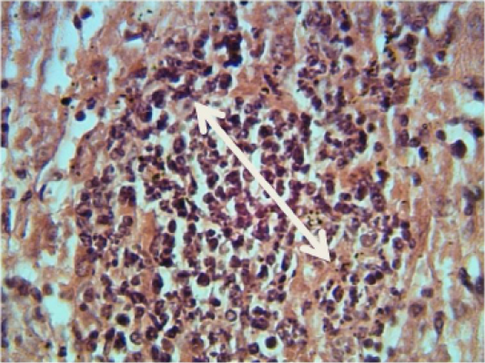
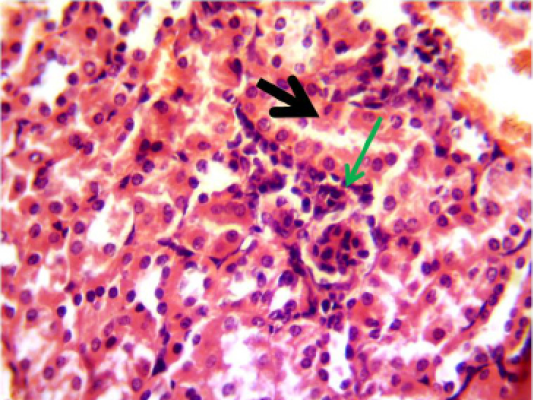
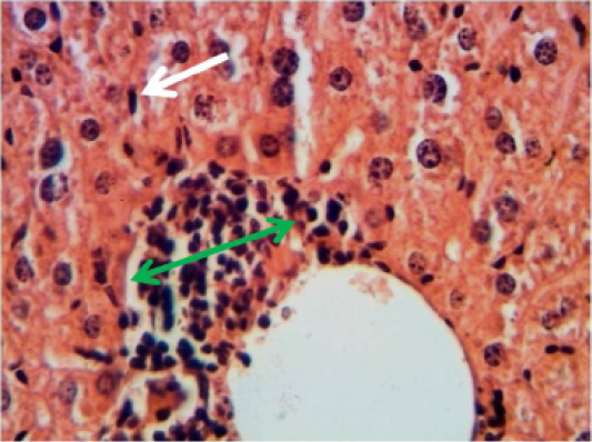
Histopathological section in the liver of animal at 6 weeks post treatment with sumithrin shows granulomatous lesion consisting from aggregation of neutrophil macrophages and lymphocytes in the liver paranchyma white arrow (H and E stain 40X)
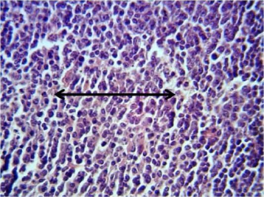
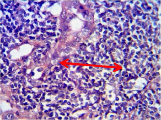
Histopathological section in the spleen of animal at 6 weeks post treatment with sumithrin shows inflammatory cells infiltration in the red pulp with depletion of white pulp black arrow (H and E stain 40X)
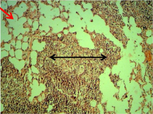
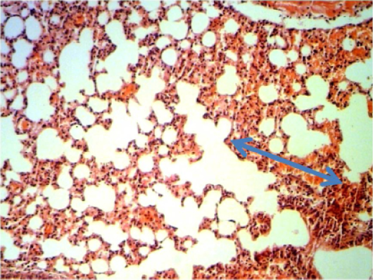
Histopathological section in the lung of animal at 6 weeks post treatment with sumithrin shows multiple abscess (dead and live neutrophils) black arrow with emphysema red arrow in the lung parenchyma with emphysema (H and E stain 10X).
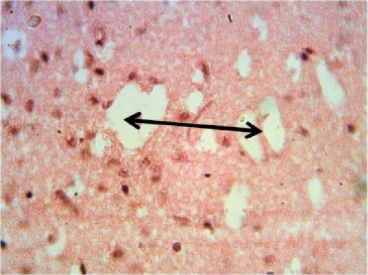
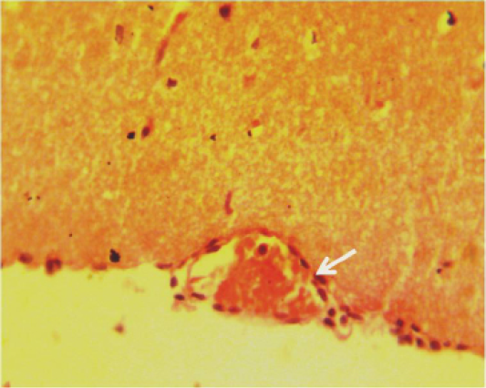
Histopathological section in the brain of animal at 6 weeks post treatment with sumithrin shows multiple irregular spaces in the brain parenchyma (spingioss) black arrow (H and E stain 40X)
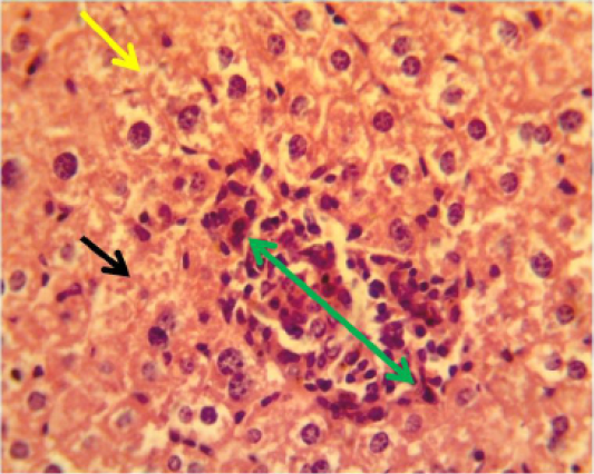
Histopathological section in the liver of animal feeding B-glucan at 6 weeks post-adminstration with sumithrinshows granulomatous lesion green arrow in the liver parenchyma with congested dilated sinusoids yellow arrow and vacuolar degeneration black arrow of hepatocytes (H and Estain 40X)
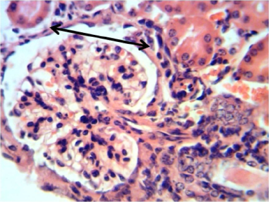
Histopathological section in the kidney of animal feeding B-glucan at 6 weeks post-adminstration with sumithrin shows vacuolar degeneration black arrow and enlargement of epithelial cells of renal tubules wth mononuclear cells infiltration around glomerula green arrow (H and Estain 40X)
Histopathological section in the lung of animal feeding B-glucan at 6 weeks post-adminstration with sumithrinshows hemorrhage in alveolar spaces and congested inter alveolarcapileries blood vessels blue arrow (H and E stain 40X)
Histopathological section in the brain of animal feeding B-glucan at 6 weeks post-adminstration with sumithrin shows inflammatory cells in the lumen of congested blood vessels in pia matter white arrow (H and Estain 40X)

Histopathological section in the liver of animal feeding B-glucan shows mononuclear cells aggregation in portal area around proliferation of bile duct and portal blood vessels white arrow (H and E stain 40X)
Histopathological section in the liver of of animal feeding B-glucanshows mononuclear cells aggregation in one side of dilated central veins green arrow with proliferation of kupffer cells white arrow )H and E stain 40X)
Histopathological section in the spleen of animal of feeding B-glucan shows moderate hyperplasia of white pulp red arrow (H and E stain 40X)
Histopathological section in the kidney of of animal feeding B-glucan shows increase thickness of glomerular wall due to fibrosis and mononuclear cell infiltration black arrow (H and E stain 40X)


