The Journal of Advances in Parasitology
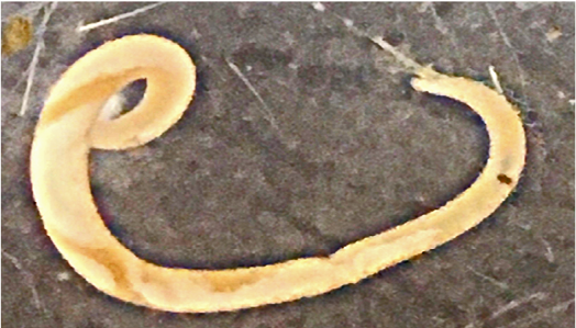
Live Dujardinascaris waltoni isolated from the stomach lumen of Alligator mississippiensis.
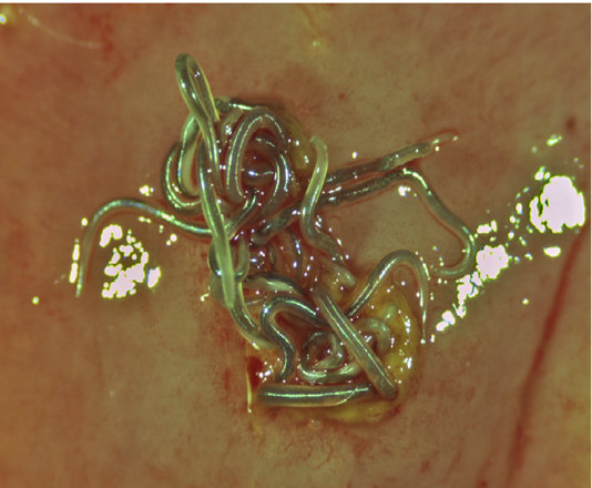
Superior view of a group of live Ortleppascaris antipini on a freshly excised portion of the stomach wall of Alligator mississippiensis. The nematodes are embedded in the mucosa at a focal nodule approximately 4 mm in diameter.
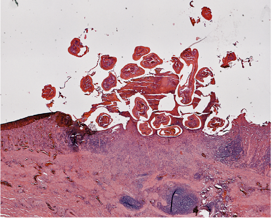
Section of the stomach wall of Alligator mississippiensis showing a lesion about 4 mm in diameter, housing multiple Ortleppascaris antipini.
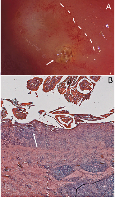
Mucosal response to the nematode invasion. A) Photograph of excised stomach wall of Alligator mississippiensis; the embedded Ortleppascaris have been removed from the lesion. The lesions (arrow, right) caused a penumbra of mucosal damage that was clearly visible (dashed lines); B) Section of the stomach wall of Alligator mississippiensis showing the gastric mucosa invaded with the Ortleppascaris. The parasites invaded the mucosa and submucosa, triggering a granulomatous inflammation response and a marked eosinophilic necrosis (arrow).
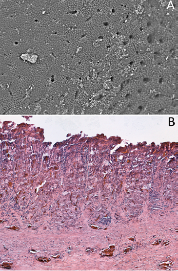
Gastric glands and pits in Alligator mississippiensis. A) Scanning electron micrograph of the surface of the stomach mucosa. This region of the stomach, which was free of parasites, shows a uniform mucosa and regular pattern of gastric pits; B) Section of the stomach wall showing the presence of “typical” vertebrate gastric pits and glands.
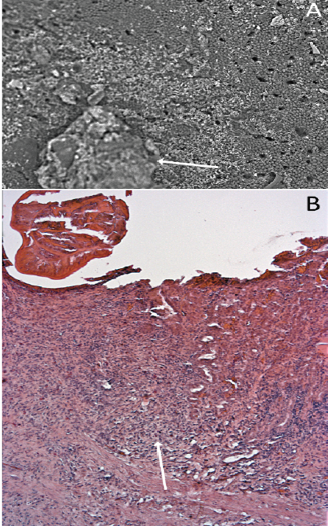
The influence of nematode infection on the gastric glands and pits of Alligator mississippiensis. A) Scanning electron micrograph showing the stomach wall of Alligator mississippiensis. The Ortleppascaris infection (arrow) has produced a central area of necrosis and a penumbra of mucosal damage; B) Section of the stomach wall of Alligator mississippiensis showing the loss of the gastric pits in the area of the Ortleppascaris invasion (arrow).


